Intense Module 1
Introduction
EXPLANATORY NOTES
In Āyurveda the term Parīkṣā is used for examination. In the present context the Parīkṣā is used for the clinical examination of the patients. There are various occasion in the Pañcakarma cikitsā when the examination of the patient’s and its disease condition is done. They are used for assessment of the various factors like screening, diagnosis and monitoring of the health condition. These factors are summarized in the following flowchart.
The Trividha parīkṣā are the three methods of clinical assessments of Patient’s profile. The three aspects that are under its umbrella are –
- Darśanaa parīkṣā
- Sparśana parīkṣā
- Prashana parīkṣā
Darśanaa Parīkṣā (visual imaging)
Darśanaa means ‘to see’ any examination done through the sense of vision is included in the Darśanaa parīkṣā. It is the observation and inspection in clinical examination through naked eye or with the help of specialized equipments. It begins at the first point of eye contact with a patient and is continued throughout the clinical encounter. It is more goal-oriented and it is limited to what one can observe visually while examining specific surface of the body parts like skin, eyes or ears etc.
Clinical significance of Visual inspection in clinical encounters –
General survey – The general survey of patients is observed through visual inspection. This provides following diagnostic clues about various qualities and potential problems of the patients like
- Patient’s medical, social, and emotional situation through clothing, jewelry, tattoos, grooming, hygiene.
- Stature and habitus – Dwarfism, pseudohypoparathyroidism, Turner’s syndrome, or prepubertal steroid therapy, Marfan’s syndrome etc.
- facial features and expression, signs of emotional distress (fidgeting excessively, exhibiting generalized psychomotor slowing, or crying, inappropriate eye contact), mannerisms
- Gait
- Posture and decubitus -. Alteration in healthy, upright posture may characteristic due to Parkinson’s disease, stroke, or cerebellar abnormalities. Decubitus ( observed posture of the patient in bed) may suggest abdominal disease, peritonitis, renal colic etc.
- Illness and severity of disease – healthy, unhealthy, weak, frail etc.
- level of consciousness – awake, alert, or somnolent
- Guarded movements (sign of pains)
- Diaphoresis
- Respiratory distress – tripoding, using accessory muscles of respiration.
- Skin lesions, abnormal fat distribution, muscle atrophy etc.
- Motor activity
- Reactions to the questions asked during the exam.
- Ancillary information regarding the patient’s social supports, interests, lifestyle, interpersonal dynamics etc can be obtained from the inspection.
Organ-specific observation – In this a detail visual physical examination of each organ system is done. In this certain examination aids are also used. The various aspect cover under this is to assess normal conditions, deviations, color, size, location, movement, texture, symmetry of each organ system.
The various tools used in organ specific examination are –
- Examination Light – It is focused on the body part to be examined.
- Laryngeal Mirror – used to exam the larynx and other areas of the throat. The laryngeal mirror reflects the inside of the mouth and throat for the physical examination. It may be used to visualize the throat for the application of anesthesia or to remove tissue from the mouth also.
- Nāsāl Speculum – inserted into the nostril to assist the physician with the visual inspection of the lining of the nose, nasal membranes and septum.
- Otoscope – allows the physician to view the ear canal and tympanic membrane. The otoscope has a magnifying lens, light and cone-shaped insert to examine the inner ear.
- Ophthalmoscope – used to examine the interior structures of the eye. The ophthalmoscope has a light, magnifying lens and opening for the physician to view the eye.
- Penlight – provides additional light for the physician to examine a specific area of the patient’s body. The penlight is typically used to examine the eyes, nose and throat.
- Percussion Hammer – used to test neurologic reflexes. The head of the instrument is used to test reflexes by striking the tendons of the ankle, knee, wrist and elbow.
- Tongue Depressors – used to depress the tongue of a patient to examine the mouth and throat during a physical examination.
Skin examination – The inspection is the main diagnostic component in the skin examination. It includes inspection of all anterior, posterior, and lateral body surfaces and mucous membranes including the hair, scalp, mastoid processes, posterior auricles, external auditory canals, nares, axilla, nails, palpebral conjunctiva, oral mucosa, inferior aspects of the breasts, skin underlying a pannus, surfaces of genitals, vaginal mucosa, and gluteal cleft. The color, [hypo- or hyper-pigmentation, pallor (palpebral conjunctiva, palms, soles, and nailbeds), cyanosis (nailbeds, lips, and perioral), and jaundice (sclera, skin, and mucous membranes)], degree of hydration (i.e. dryness or oiliness), turgor and texture of the skin are noted in it.
Aided Visual examination – In this type of darśanaa parīkṣā certain aids are used which enhance visual perception and allows looking beyond the limitation of naked eyes. These are –
Body Scans –
- X-ray -Uses x-radiation to take images of dense tissues inside the body such as bones or tumours.
- Ultrasound – A scan that uses sound waves to create images from within the body.
- CT -The Computer Tomography (CT) scan takes a number of x-rays to make a 3D image of an affected area.
- EUS – The Endoscopic Ultrasound Scan (EUS) uses a tube-like instrument called an endoscope with an ultrasound scanner attached. This is put inside the body to look inside the gut to investigate GIST tumours.
- PET – The Positron Emission Tomography (PET) scan shows up changes in tissues that use glucose as their main source of energy – for example, the brain or heart muscle. It involves an injection of a very small amount of a radioactive drug into the body. The drug travels to places where glucose is used for energy and shows up cancers because they use glucose in a different way from normal tissue.
- MRI -Magnetic Resonance Imaging (MRI) uses magnets to create an image of the tissues of the body.
- Bone scan -Uses radioactive chemicals called radionuclides which are injected, swallowed or breathed into the body, to take images of bones.
Microscopic studies –
- Tissue Analysis – histological study cells and tissues.
- Genetics Study – genetic abnormality, regeneration and tissue death.
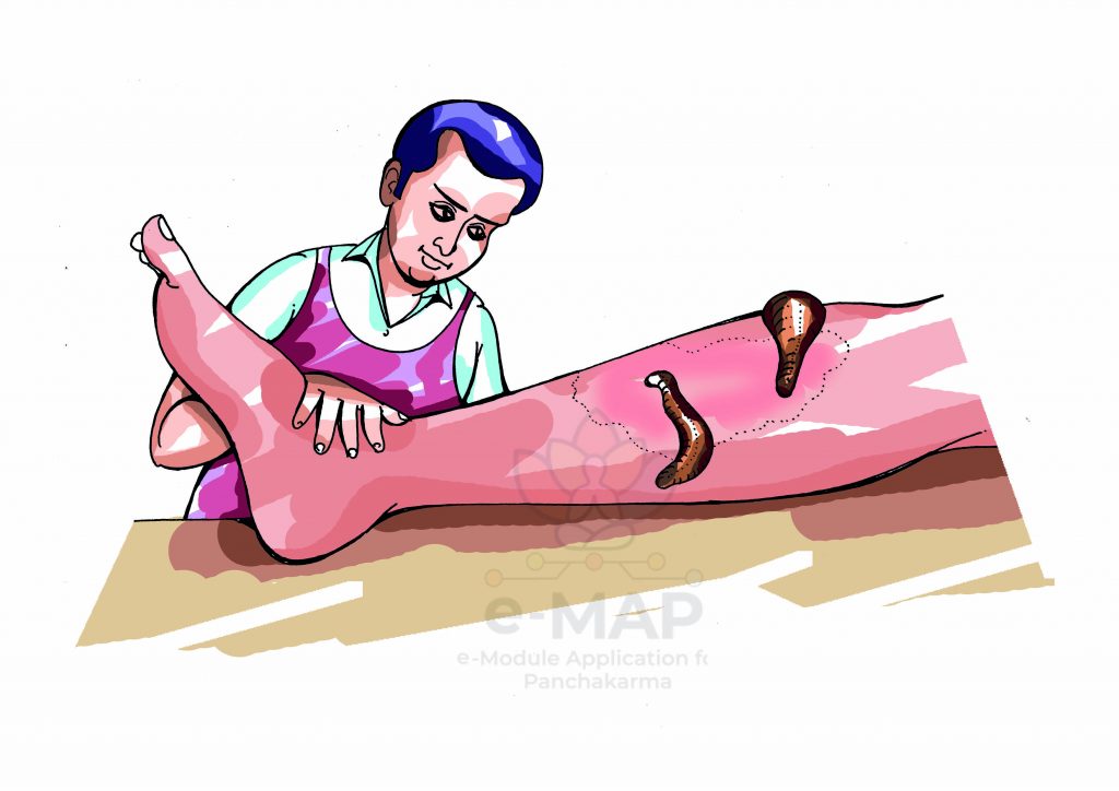
Sparśana Parīkṣā (tactile imaging)
The Sparśana means the sense of touch, any information that is obtained by the tactile perception is termed as the Sparśana parīkṣā . The most common example of Sparśana Parīkṣā is the use of thermometer to assess the body temperature. The palpation and Percussion are included in Sparśana Parīkṣā. The various aspect of sparśana parīkṣā are –
Palpation – It requires to touch the patient with different parts of hands, using varying degrees of pressure. The examples of condition that requires palpation are pulse diagnosis, abdominal distension, cardiac thrills, fremitus, various hernias, joint dislocations, bone fractures, and tumors etc .
Types of Palpation with their clinical significance –
Manual Palpation
It is done with the bare hands with or without support of three-dimensional (3D) digitizing. It is used for identification of painful areas, positioning of particular pieces of equipment (electromyography electrodes, auscultation, external landmarks used in body surface scanning), measurements of morphological parameters (e.g., limb length) etc. The two types of manual palpation are –
- Light palpation – it is used to feel surface abnormalities with finger pads, using the lightest touch possible, to assess texture, tenderness, temperature, moisture, elasticity, pulsations, and masses.
- Deep palpation – it is used to feel internal organs and masses for size, shape, tenderness, symmetry and mobility with firm deep pressure.
Virtual palpation
It is done through the 3D computer models like medical imaging. The example of virtual palpation are –
- Elastography – Elastography is a medical imaging technique to determine the elastic properties and stiffness of soft tissue for diagnostic information about the presence or status of disease ( in liver, breast, thyroids, prostate, musculoskeletal, brain, kidney etc). The Elastography is also used for guiding biopsies. Types of Elastography includes –
- Ultrasound elastography – Quasistatic elastography (strain imaging), Acoustic radiation force impulse imaging (ARFI), Shear-wave elasticity imaging (SWEI), Supersonic shear imaging (SSI), Transient elastography etc.
- Magnetic resonance elastography (MRE)
- optical coherence tomography
- Computerized palpation – In this type of imaging sense of touch is converted into a digital image. It is also called “Mechanical imaging” or “Stress imaging”. In this the probe of the device with a pressure sensor slightly deforms soft tissue just like human fingers and detect changes in the pressure pattern on tissues.
Percussion – It involves tapping fingers or hands quickly and sharply against parts of the patient’s body to help locate organ borders, identify organ shape and position, and determine if an organ is solid or filled with fluid or gas.
- Direct percussion – to reveals tenderness
- Indirect percussion – to elicits sounds that give clues to the makeup of the underlying tissue.
Clinical significance of Percussion –
- used to diagnose pneumothorax, emphysema and assess the respiratory mobility of the thorax.
- used to find organomegaly
- used to determine tissue health or pathology.
- Tympanitic, drum-like sounds heard over air filled structures. during the abdominal examination.
- Impaired resonance, dull sound in mass or consolidation.
- Percussion induce pain indicating underlying pathology.
Prasana Parīkṣā (medical interview)
The Prasana parīkṣā in the Āyurveda is medical interview. It is a purposeful conversation for diagnosis and planning therapeutic measure. The clinical hypothesis generated through the medical interview is far most cost effective than any other clinical or diagnostic techniques. It develops a positive relationship with the patient initiate the process of patient care. The various information that are collected through the medical interview are –
Types of Medical interview –
- The Problem-Oriented Interviews – It is basically directed towards the patient’s interest in seeking a solution for his current health issue. These are performed in regular O.P.D units.
- Health Promotion Interviews – In these all the past and present events of the patients life are assessed to establish the health risk and early evidences of the disease of patients. These type of interviews are often performed in the routine health checkups.
Clinical significance of Medical interview –
- Suggest diagnosis – It suggest the probable diagnosis though the chronological record of patient’s health status described by himself.
- Disclose patients profile – It provides the information regarding the patient’s behavior , emotional concerns, mental status, Physical, psychological and social profile.
- Initiate therapeutic measure – The conversation with the patient develops the interpersonal relationship and trust. This paves the way for therapeutic goals.
- Act as a mean of Psychotherapy – The medical interview assist the patient in relieving his helplessness in disease condition and act as a mean of Psychotherapy.
The Trividha roga parīkṣā are the methods to analyze the disease condition. The component of Trividha roga parīkṣā are –
- Aaptopadeśa
- Pratyakṣam
- Anumāna
Aptopadesh Pareeksha – (Evidence-Based Clinical Practice Guidelines)
In Āyurveda Aptopadeśa is the term used to describe the guidelines from the authoritative. The Aptas are most reliable and truthful on that particular topic. It is like expert testimony. The main characteristics features of Apta are –
रजस्तमोभ्यां निर्मुक्तास्तपोज्ञानबलेन ये|
येषां त्रिकालममलं ज्ञानमव्याहतं सदा||१८||
आप्ताः शिष्टा विबुद्धास्ते तेषां वाक्यमसंशयम्|
सत्यं, वक्ष्यन्ति ते कस्मादसत्यं नीरजस्तमाः ||१९|| ch.soo.11/17-19
तत्राप्तोपदेशो नामाप्तवचनम्|
आप्ता ह्यवितर्कस्मृतिविभागविदो निष्प्रीत्युपतापदर्शिनश्च|
तेषामेवङ्गुणयोगाद्यद्वचनं तत् प्रमाणम्|
अप्रमाणं पुनर्मत्तोन्मत्तमूर्खरक्तदुष्टादुष्टवचनमिति ch.vi.4/4
स्व कर्मण्य अभियुक्तो यः राग द्वेष विवर्जीताः |
निर्वैरः पूजितः सिद्धिः आसो ज्ञेयः स दादृशः ||
आप्तः तु यथार्थ वक्ता | ( संख्यकारिका)
- Raja Tamobhyāma Nirmuktama, Nishpreeti Uptaap Darshina – They are liberated at spiṛtual level from any presumptive notion. They are not bias.
- TriKālāmaGyan – They have a complete knowledge of the subject in terms of time span (past, present and future)
- Amala Gyan, Yathaartha Vakta, Gyaayate Anene Iti Pramaanam, – Their knowledge is complete truth, reliable and treated as evidence.
- Avayahata Sada, TeshāmaVakya Asanshayāma – None of their fact is distorted or doubtful.
- Shishta, Vibuddha – They are disciplined,well behaved, highly intellectual.
- Avitarka Smruti, Vibhaga Vido – Their have sound retention power and have keen observation.
- Swa Karmanyoabhiyukta – They are dedicated to their work.
- Nirvarihi – They do not have antagonist.
Thus Aptopadesh implies the evidence or data collected through the patient’s examination are analyzed on the basis of pre-existing set of categories or clinical practice guidelines agreed upon by the medical profession to designate a specific condition. The clinical practice guidelines are statements that include recommendations intended to optimize patient care.
Pratyaksha Pramāṇa
In Āyurveda the Pratyaksha Pramāṇa is the evidence obtained as a resultant outcome of the cumulative action of Atma, Indriya, mana and artha. The diagnosis is made on the basis of the evidence obtained through the physical examination of the patients and various diagnostic test carried on the patient. In Āyurveda this is obtained by the use of all the four senses – Shadba, Sparśa, Roopa, Gandha. In present times the Rasa can also be analyzed indirectly. On the basis of Indriya (sensory perception) the pratyaksha are classified as –
- Sabda pratyaksha Pramāṇa
- Sparśa pratyaksha Pramāṇa
- Rūpa pratyaksha Pramāṇa
- Rasa pratyaksha Pramāṇa
- Gandha pratyaksha Pramāṇa
Sabda pratyaksha Pramāṇa – The auditory perception used in the examination of the patient is termed as Sabda Pratyaksha Pramāṇa. Auditory perception could be defined as the ability to receive and interpret information that reached the ears through audible frequency waves transmitted through the air or other means. Through the sense of auditory perception various clinical condition like deafness, Wernicke’s aphasia (inability to understand language), auditory agnosia (inability to recognize a heard object), amusia (unable to recognize or reproduce tones or musical rhythms), tinnitus (to hear a constant ringing), hallucinations (schizophrenia, paracusis of Willis) etc, can be identified. The various other examples of Auditory perception are Antarkoojana, Sandhisputhana, swara visesha, sirpagatha sabdha etc. In modern medicine the Sabda Pratyaksha Pramāṇa can be achieved through Auscultation. The Auscultation is listening of the sounds from the internal organs of the body. Majorly the sounds of circulatory, respiratory and gastrointestinal systems (heart, lungs and bowel sounds) are examined.
The various other mode of achieving Sabda pratyaksha Pramāṇa are –
- Stethoscope – used to listen body sounds including the sounds of the heart, lungs and intestines.
Method of auscultation by stethoscope – Maintain a quiet environment. Make sure the area to be auscultated is well exposed as the cloth or covering can interfere with the sound. Warm the stethoscope head with the palms and focus attention. Then place the diaphragm firmly against the patient’s skin, using enough pressure to leave a slight ring on the skin afterward for high pitched sounds like first (S1) and second (S2) heart sounds .For low pitched sounds like third (S3) and fourth (S4) heart sounds, the bell is lightly held against the patient’s skin, just hard enough to form a seal because holding the bell too firmly can causes the skin to act as a diaphragm and obliterating low-pitched sounds. Then concentration is made to listen and identify the characteristics of one sound at a time.
- Sphygmomanometer – used to measure a patient’s blood pressure. The sphygmomanometer is composed of an inflatable rubber cuff, a bulb that inflates and releases pressure from the cuff, and use of a stethoscope to listen to arterial blood flow in the patient.
- Audioscope – used to screen patients for hearing loss. The audioscope is placed in the patient’s ear and makes a series of tones which the patient can respond to.
- Tuning Fork – used to test a patient’s hearing. The physician strikes the prongs causing them to vibrate and produce a humming sound. Then the prongs are placed next to the patient’s skull, near the ear, with the patient describing what they heard. The physician may order additional tests depending on the results of this hearing test.
Sparśa pratyaksha Pramāṇa – The details of sparśa pratyaksha Pramāṇa are discussed in the Sparśana Parīkṣā of Trividha Rogi Parīkṣā in the previous section of the unit.
Rūpa pratyaksha Pramāṇa – The details of Rūpa pratyaksha Pramāṇa are discussed in the Darśanaa Parīkṣā of Trividha Rogi Parīkṣā in the previous section of the unit.
Rasa pratyaksha Pramāṇa – In Āyurveda the Rasa pratyaksha from one’s own sense is discouraged but example of inference drawn from rasa pratyaksha from other organism are present. Basically the taste perception is due to specific chemical structure of the substance that interact with the taste receptors (gustatory system). Taste perception or determination of the flavor is a combined resultant of Gustatory function (upper surface of the tongue and the epiglottis), smell perception and Trigerminal nerve stimulation. The final conclusion is drawn by the gustatory cortex in the brain. The six basic taste which are identified are –
- Madhura – sweet
- Amla – sour
- Lavana – salty
- Katu – spicy
- Tikta – bitter
- Kaṣāya – Astriṇgent
The sweet sour and bitter taste are indentified by the binding of molecules to G protein-coupled receptors on the cell membranes of taste buds (chemical structure is identified), whereas saltiness and sourness are identified when OH– or H+ ions enters taste buds (this basically identifies the pH of the substance). The other perception that are drawn are the smell (along with the olfactory epithelium of the nose), texture (by mechanoreceptors), temperature (thermoreceptors), pungency (Chemesthesis)
The taste perception in present times are achieved through varieties of supportive like –
- pH calculators –The pH calculators identifies the pH on scale from 0 to 14. It tells how acidic or alkaline a substance is. More acidic solutions have lower pH (less than 7). More alkaline solutions have higher pH (greater than 7). Neutral usually have a pH of 7.
- Glucometres – based on the oxidation of glucose to gluconolactone catalyzed by glucose oxidase or glucose dehydrogenase enzyme.
- Glucose test strips for urine – Work on the principle of colorimeters.
- Blood alcohol sensors
- Various types of Chromatography – to identify the presence and relative proportions of the analytes in a test product (blood, sweat, Cerebrospinal fluid, saliva, tears, urine, plasma, semen, breast milk, Chylous effusion, drainage fluids, pericardial fluids, peritoneal fluids, etc).
- Mass spectrometry
Gandha pratyaksha Pramāṇa – The Gandha pratyaksha Pramāṇa is the olfactory perception used for the diagnosis of the condition of the patient. There are various theories in Chinese medicine regarding the odor of the body like the change in odor of the body may be due to –
- Disturbance in gut flora
- Due to stress or anxiety – smoky odor due to excessive cortisol
- Liver and gallbladder disorders – Rancid body odors
- Spleen diorder – sweet smelling body odor
- Lung or large intestine disorder – Rotten odor
- Kidney disorder – Putrid body odors, similar to the smell of ammonia or sea water
In modern medicine it is considered that some diseases have breath print that help them to identify them. The reason that is assumed for this fact is the disease leads to development of new and different biochemical processes in the body, which lead to the production of different volatile molecules when these reaches to the lungs and be released in exhaled breath or released in the urine and sweat, they cause characteristics smell. The few disease on which researches of breath prints are undergoing are –
- Lung cancer – An invention called “NaNose”—a breathalyzer-type device developed by an Israeli company—is up to 90 percent accurate at diagnosing lung cancer; the device detects a special “odor” emitted by the cancer cells. Parkinson’s disease, kidney failure, multiple sclerosis, Crohn’s disease and other cancers are also elevated with accuracy rate is at 86 percent,
- Preeclampsia
- Kidney disease – Ammonia on the breath is a sign of kidney failure
- Diabetes – fruity smell
- Breast cancer
- Melanoma
- Liver disorder – odor like raw fish is the characteristics feature of liver
- Gum disease
- Infectious mononucleosis – sour breath
- Trimethylaminuria – rotting fish, urine, days-old garbage or rotten eggs
- Schizophrenia – Bad breath
- Maple syrup disorder – Maple syrup
The various disorders of smell indentified by the patients himself are –
- Anosmia – complete loss of ability to smell
- Hyposmia – Reduced ability to smell
- Parosmia – distorted odor perception
- Phantosmia – olfactory hallucination
The causes of the smell disorders includes illness such as upper respiratory infection, injury, polyps in the nasal cavities, sinus infections, hormonal disturbances, dental problems, exposure to certain chemicals such as insecticides and solvents, some medicines, and radiation due to head and neck cancers.
Anuman Parīkṣā
The Anumāna Parīkṣā is inferential diagnosis. Various facts of clinical condition and differential diagnosis of various conditions are analyzed to establish an inferential diagnosis.
The few examples of inferential diagnosis are Power of Agni is analyzed by the process of digestion and metabolism, exercise endurance capacity decides the strength of that person. The prime factor that establish the inference is on the differential diagnosis. Differential diagnosis is a hypothetico-deductive method used to distinguishing between various clinical condition based on the clinical feature. Various approaches are used in differential diagnosis like process of elimination, estimating Probabilities on the basis of evidence generated though presenting symptoms, patient history, and medical knowledge, diagnostic measures etc.
Ṣaḍvidha Parīkṣā of a patient is a method of examination of patient for the diagnosis of the its disease condition and assessment of physical condition. The six method of the Ṣaḍvidha Parīkṣā are –
- Sabda Parīkṣā
- Sparśa Parīkṣā
- Rūpa Parīkṣā
- Rasa Parīkṣā
- Gandha Parīkṣā
- Prashna Parīkṣā
The details of the above six method of examination is described in previous section of the unit in Pratayaksha Pramāṇa and medical interview. .
The Aṣṭavidha Parīkṣā are the method of clinical examination that is used to assess the Rogi Roga Bala, and Vyādhi Viniścaya (patient’s health, diagnosis and prognosis ). The eight aspects of health are –
- Nāḍi Parīkṣā (Pulse diagnosis)
- Mala Parīkṣā (Stool diagnosis)
- Mūtra Parīkṣā (Urine test diagnosis)
- Jivhā Parīkṣā (Tongue diagnosis)
- Śabda Parīkṣā (Voice analysis diagnosis)
- Sparśa Parīkṣā (Dermatological diagnosis)
- Dṛk Parīkṣā (Eye Examination)
- Ākṛti Parīkṣā ( Face and General appearance Examination)
Nāḍi Parīkṣā (Sphygmology, Pulsology)
The Nāḍi Parīkṣā is the pulse examination. It is a unique concept of Āyurveda. From the pulse examination various assessment of patient’s health can be obtained. The Nāḍi at Aṅguṣṭamūla base of thumb is analysed to estimate the quantum of Tridoṣa in the body, establish diagnosis and assess prognosis in Roga and Rogi Parīkṣā. It is considered that the three Nāḍi’s – Vāta, Pitta and Kapha lies under index, middle and ring finger respectively.
Method of Nāḍi Parīkṣā –
- The ideal time for Nāḍi assessment is early morning with empty stomach for Prakṛti analysis (physiological profile).
- The Nāḍi Parīkṣā is contraindicated immediately after bath, immediately after having food, after massaging, hungry, thirsty and while sleeping.
- The patients and the assessor both should have mental stability and peace of mind.
- There are eight Nāḍi sites two are Hastadvayagata Nāḍis, which are located at the end of Prakoṣṭha (fore arm) and three inches below the Maṇibandha (wrist). Two are Pādadvaya Gata Nāḍis; these are located below Gulpha (ankle) around three inches level. Two are Kaṇṭa Pārśva Nāḍies. These are located at the root of the neck in both sides around one inch level. Another two are Nāsāmūlagata Nāḍis, which are located around one inch at Nāsāmūlam (root of nose). The radial pulse is preferred due its superficial position and ease of palpability.
- To determine Āgantuka Jvara, Tṛṣṇa (thrist), Āyāsa (dyspnoea), Maithunasaṅklamana (fatigue due to copulation), Bhaya (fear), Śoka (sorrow), Kopa (anger), Kaṇṭa Nāḍi should examined.
- To determine Mṛtyu (death), Kāma (desire), Netraroga (eye disorder) Śirovyatha (head ache), Śravaṇa (ear) Mukharogas (mouth), Nāsā Nāḍi should be examined.
- The right hand’s Nāḍi is assessed in male patients and for females left hand Nāḍi is assessed.
- The physician should support the patient right palm and forearm with his left hand, then the physician should examine the patient’s Aṅguṣṭha Mūla (root of thumb i.e., below the thumb in the wrist region), with his Dakṣiṇa Kara Aṅgulītraya (Right hand’s middle three fingers).
- The elbow of the patients is slightly flexed towards left and the wrist should be slightly bended to the left with distended and dispersed fingers.
- Nāḍi is assessed in three repeated attempts by releasing and applying pressure on Doṣic spots.
- Vāta Nāḍi is assessed on the index finger, Pitta Nāḍi is assessed in middle fingers and Kapha Nāḍi is assessed on ring finger, Sannipata Nāḍi is vibrated all the three fingers.
- After the Nāḍi examination the physician should wash his hands.
The various Rthyms of the Nāḍi as per the text are –
- Vātaja Nāḍi – Snake and leech
- Pittaja Nāḍi – Crow lark and frog
- Kaphaja Nāḍi – Swan, Pigeon and cock
- Vāta Kaphaj Nāḍi – Snake and swan
- Pitta- Kaphaja Nāḍi – Monkey and swan
- Vāta Pittaja Nāḍi – Snake and frog
- Saṃnipātaja Nāḍi – Wood pecker
- Jvara – Gambhīra Uṣṇa And Vegavati
- Kāmakrodha – Vegavati (Rapid)
- Cinta and Bhaya – Kṣīṇa (Weak)
- Mandāgni – Manda (Slow)
- Rakta Doṣa – Uṣṇa, Gurvī (Heavy) And Sāma
- Āma– Gambhīra
- Diptāgni – Laghu And Vegavan
- Kṣudhita – Cañcala (Unstable)
- Tṛpta – Sthira (Stable)
- Asādhya Vyādhi – Kampana (Vibration) And Spandana (Pulsation)
- Vāta Jvara – Capala (Unstable), cold on touch
- Pitta Jvara – Rapid, straight and of long duration
- Kapha Jvara – Slow, stable, cold and sticky
- Vāta Pitta Jvara – Somewhat Vakra, Capala and Kaṭhina
- Kapha Vātaja – Manda (Slow)
- Pitta Kapha – Sūkṣma, Śītala And Sthira
Pulse examination – The tactile arterial palpation of the heartbeat is termed as pulse examination. The pulse count is equivalent to heart rate. The pulse examination is conducted on the artery that are near to the body surface like – carotid artery on neck , radial artery on wrist , femoral artery in groin, popliteal artery behind the knee, posterior tibial artery near the ankle joint and dorsalis pedis artery on foot. The pulse examination is conducted using the three fingers . The reason behind it is the finger closest to the heart is used to occlude the pulse pressure, the middle finger is used get a crude estimate of the blood pressure, and the finger most distal to the heart (usually the ring finger) is used to nullify the effect of the ulnar pulse as the two arteries are connected via the Palmar arches (superficial and deep).
The various assessments of the pulse examination are –
- Determination of systolic blood pressure.
- The pulse deficit (difference between heart beats and pulsations at the periphery) is determined by simultaneous palpation at the radial artery and auscultation at the PMI, near the heart apex. It may be present in case of premature beats or atrial fibrillation.
- Determination of pulse rate – The pulse rate determines the health status of a subject. The lower limits ascertain the good condition but severely slow Heart rate (bradycardias) is a sign of weakness, loss of energy and fainting.
Normal pulse rates at rest, in beats per minute (BPM) | |
Newborn (0–3 months old) | 99-149 |
Infants (3 – 6 months) | 89–119 |
Infants (6 – 12 months) | 79-119 |
Children (1 – 10 years) | 69–129 |
Children over 10 years & adults, including seniors | 59–99 |
Well-trained adult athletes | 39–59 |
- Determination of Pulse rhythm and force – Normally a healthy pulse is regular in rhythm and force. The cause of irregularity in the pulse is pathologic.
Irregular pulse | sinus arrhythmia, ectopic beats, atrial fibrillation, paroxysmal atrial tachycardia, atrial flutter, partial heart block |
Regularly irregular pulse | pulsus bigeminus, second-degree atrioventricular block |
Irregularly irregular pulse | Atrial fibrillation |
- Estimation of Form – A quick rise and fall in pulse (pulsus celer) is seen in aortic regurgitation. A slow rise and fall in pulse (pulsus tardus) is seen in aortic stenosis.
- Comparison – An inequality between the left and right radial pulse or upper and lower extremities is observed in coarctation of aorta, aortitis, dissecting aneurysm, peripheral embolism etc
- Arteriosclerosis – Arteriosclerosis can be determined from a palpable artery after flattening by digital pressure.
- Dicrotic pulse (two palpable pulsations, the second of which is diastolic and immediately follows the second heart sound) due to low cardiac output and high systemic vascular resistance.
- Pulsus alternans – A pattern of a strong pulse followed by a weak pulse over and over again in progressive systolic heart failure.
- Pulsus bigeminus groups of two heartbeats close together followed by a longer pause. The second pulse is weaker than the first, indicates hypertrophic obstructive cardiomyopathy.
- Pulsus bisferiens ( two beats per cadiac cycle, both systolic), indicated aortic valve diseases
- Pulsus tardus et parvus, also pulsus parvus et tardus, slow-rising pulse and anacrotic pulse, is weak (parvus), and late (tardus) indicates aortic valve stenosis.
- Pulsus paradoxus (radial pulse not detected during the inspiration) indicates cardiac and respiratory conditions of emergency.
- Tachycardia( an elevated resting heart rate)
- collapsing pulse (sign of hyperdynamic circulation).
For further detail study on Nāḍi Parīkṣā following papers may be referred –
- Kumar PVG, et al., Traditional practices and recent advances in Nāḍi Parīkṣā: A comprehensive review, Journal of and Āyurveda Integrative Medicine (2017), ttps://doi.org/10.1016/j.jaim.2017.10.007
- Prasad GP, Bharati K, Swamy RK. Some important aspects of Nāḍiparīkṣā from bāsavarrājīyam. Anc Sci Life. 2004;24(1):27–29.
- Tripathi AK, Singh RH. Role of Nāḍi Pariksa in the diagnosis of Udara Roga. Anc Sci Life. 1994;13(3-4):248–252.
Mala Parīkṣā
The Mala Parīkṣā is the stool examination of Āyurveda. The various factors examined through Mala Parīkṣā are Agni (metabolism), Doṣa (Physiological profile), Vyādhi (Pathological state), Krimi (worms infestation). Basically the motive of stool analysis is to identify certain conditions of digestive tract like infection, malnutrition, cancer etc. The various characteristics that are observed during Mala Parīkṣā are quantity or volume, shape, color, odor, consistency, presence of mucous, presence of blood, etc.
The various examination used for Mala Parīkṣā are –
- Sabda parīkṣā – Patients self-description of the complaints come under the scope of Śabda Parīkṣā. The various informations provided by the patients are –
Quantity and frequency – Once or twice a day frequency is considered normal and the total quantity of Mala should not exceed Sapta Anjali of the individual patient. The Alpa quantity is indicative of Samnnipātaja Jvara, Pāṇḍu, Vātaja Atisāra and Purīṣavaha Stroto Duṣṭi. Atipravarti and Atimātra is observed in Paittika Atisāra, Āmātisāra Sannipātika Atisāra, Asādhya Atisāra, Saṅga (less frequency) is observed in Vātika Atisāra, Ślaiṣmika Atisāra, Pravāhika, Vātika Grahaṇī, Chidrodara etc. Muhurmuhur (repeated frequency) is observed in Vātaja Grahaṇī and Vātaja Atisāra. Muhurmuhur Bheda ( frequent loose stool ) is observed in Yakṣmā. Kṛcchra Mala Pravarti is observed in Vātaja Aśmarī, Vidvighāta, Āmāśaya and Pakvaśaya Gata Vāta. Saśabda Mala Pravarti is observed in Vātaja Arśa Āmātisāra, Vātaja Grahaṇī.
- Sparśa Parīkṣā –
Temperature – The temperature of the stool is also indicates the pathological condition like the Śītata is due Ajīrṇa and Śleṣmika Atisāra and Uṣṇata is due to Paittika Arśa.
Texture – The texture like Snigdhata (softness) is indicative of Śleṣmika Arśa and Kaphaja Arśa and Rūkṣata (roughness) is indicative of Vātaja Jvara and Vātaja Atisāra.
- Rūpa parīkṣā – The various factors that are under the scope of Rūpa Parīkṣā are –
Color – Due to various physiological and pathological conditions there is variation in color of the stool.
- ŚuklaVarṇa (white coloration) due to Śleṣmika Jvara, Śleṣmika Udararoga, Śleṣmika Arśa Śleṣmika Pāṇḍu, Śleṣmika Atisāra, śākhāśrita, Kāmala, Sahaja Arśa Jalodara, Kaphaja Gulma, Kaphaja Visarpa etc.
- Śyāva Aruṇa (reddish black coloration) due to Vātika Udara Roga, Vātika Arśa, Vātika Atisāra, Vāta Prakopa, Vātaja Gulma, Vātaja Jvara, Vātaja Pāṇḍu etc.
- Rakta Varṇa (blood red coloration) due to Kāmala, Pittaja Atisāra, Raktapitta, Sahaja Arśa Chhidrodara etc.
- Kṛṣṇaḥ Varṇa (black coloration) is due to Vāta Prakopa, Vātika Arśa Vātika Gulma, Kumbha Kāmala etc.
Consistency of the stool – It is indicative of various pathological states like – Sāndra (dense) stool is observed in Kaphaja Atisāra and Sahaja Arśa , Baddha and Vibaddha (excessive compact ) stool is observed in Udara Roga, Baddhodara, Purīṣāvṛta Vāta, Vātaja Atisāra, Vātaja Jvara, Dravya (watery liquid) stool is observed in Paittika Arśa Vātika Grahaṇī etc, Bhinna (loose ) stool is observed in Paittika Arśa Śleṣmika Grahaṇī, Kṣayaja Kāsa, Kaphāvṛta Apānavāyu, Sushka (Dry) stool is observed in Vātaja Grahaṇī and Sahaja Arśa.
Presence of mucous – Due to presence of mucous there is frothiness in stool. This help in diagnosis of condition like Arśa Atisāra, Grahaṇī of Vāta dominance.
Microscopic Examination stool – The microscopic examinations is also included under the Rūpa Parīkṣā
- Rasa parīkṣā – The Rasa Parīkṣā of the stool is indirectly done. Like use of pH strips
- Gandha Parīkṣā – The characteristic odor is the also indicative of pathological state like – Durgandhita (foul smelling) is observed in Śleṣmika Atisāra, Paittika Pāṇḍu, Asādhya Saṃnipātaja Chardi, Āma Purīṣa, Purīṣavaha Stroto Duṣṭi, Visragandhi (raw meat smell ) is observed in Paittika Arśa and Kaphaja Atisāra, Atidurgandhita ( extremely foul smelling) is observed in Paittika Arśa and Atisāra, Āmagandhi in Āmātisāra Kuṇapagandhi in Chidrodara and Sahaja Arśa Kuṇapa- Pāyu- Āma- Matsyagandhi in Saṃnipātaja Atisāra. Mahatpūtigandhi (excessive rotten smell) in Jalodara, Nirganda or Sagandha in Āgantuja Atisāra.
- Prashna parīkṣā – Through asking question various associated condition can be known in a case.These associated complaints are indicative of pathological state as Saśūlam Malapravarti ( Āmatisāra), Saśūlam and Sadāham Malapravarti (Paittika Atisāra). Parikartika ( Vātaja Atisāra), Pravāhaṇa (Pravahika), Śūla and Guḍasrāva (Jalodara), Cira Dukhāma(Vātaja Grahaṇī), Saśūlam Pravāhaṇa (Kaphaja Arśa Kaphaja Atisāra and Pravahika), Kuntana (Sāmātisāra and Viṣamāgni).
- Jala Nimajjana Parīkṣā – This is a novel method of examining the presence of Āma in stool. It is use in diagnosis of Jvara, Āmavāta, Atisāra. In this method the mala is suspended in water and its activity is observed. If the stool floats on the water it indicates the absence of Āma. If the stool sink in the water, it indicate presence of Āma.
Stool examination – A stool analysis is a series of tests done on a stool sample through microscopic examination, chemical tests, and microbiologic tests. The stool is checked for color, consistency, amount, shape, odor, pH and the presence of mucus, hidden (occult) blood, fat, meat fibers, bile, white blood cells, and sugars called reducing substances. It is done to identify diseases and functioning of the digestive tract, liver, and pancreas through enzymes (such as trypsin or elastase) evaluation in stool. It is used find causative of symptoms affecting the digestive tract, like prolonged diarrhea, bloody diarrhea, an increased amount of gas, nausea, vomiting, loss of appetite, bloating, abdominal pain, cramping and fever. Screen for colon cancer by checking for hidden (occult) blood, screen infestation & infection from parasite (pinworms or Giardia), bacteria, a fungus, or a virus. Determination for poor absorption of nutrients by the digestive tract (malabsorption syndrome) through quantitative fecal fat test. High levels of fat in the stool may be caused by as pancreatitis, sprue (celiac disease), cystic fibrosis, or other disorders that affect the absorption of fats. The presence of undigested meat fibers in the stool may be caused by pancreatitis. A low pH may be caused by poor absorption of carbohydrate or fat. Stool with a high pH may mean inflammation in the intestine (colitis), cancer, or antibiotic use. Blood in the stool may be caused by bleeding in the digestive tract. White blood cells in the stool may be caused by inflammation of the intestines, such as ulcerative colitis, or a bacterial infection. Rotaviruses are a common cause of diarrhea in young children. High levels of reducing factors in the stool may mean a problem digesting some sugars. Low levels of reducing factors may be caused by sprue (celiac disease), cystic fibrosis, or malnutrition. Medicine such as colchicine (for gout) or birth control pills may also cause low levels.
Mūtra Parīkṣā
The Mūtra Parīkṣā is the Ayurvedic assessment of urine for the diagnosis and prognosis of the disease and health status of a being. The various disease conditions where the Mūtra Parīkṣā is indicated are Jvara, Raktapitta, Prameha, Pāṇḍu, Kāmala, Aamavāta, Arśa, Aśmarī, Mūtrakṛcchra, Mūtratisāra, Mūtrasāda, Uṣṇavāta etc.
Symptoms of normal urine – The healthy human urine is Kṣāra (Alkaline), Tīkṣaṇa (Pungent), Lavaṇa (salty), resembles to the Kūpa Jala (the water of a well), Anāvila (clean , non-turbid), Apicchila (non-slimy), Viśada (non-sticky) .
Minimum volume of urine – The minimum volume of urine is four Añjali (200ml), amount lesser 200 ml indicates pathology.
- Śabda Parīkṣā – Patients self-description of the complaints come under the scope of Sabda Parīkṣā.
- Sparśa Parīkṣā – In Sparsh Parīkṣā the temperature and viscosity of the urine is analyzed as in Śītameha
- Rūpa Parīkṣā – The various information that are included in Darśana Parīkṣā are the examination that are done by naked eyes like the color, volume, transparency, turbidity etc . The microscopic examination also comes under the scope of Darśana Parīkṣā . The various appearance that are described for identification of Doṣas in Mūtra are –
- Śukla- Udakameha, Piṣṭameha , Kaphaja Pāṇḍu
- Pāṇḍur Varṇa – Vāta Doṣa
- Shwet Varṇa, Budbudabham (with bubbles) – Vāta Kapha Prakopa
- Pita – Pitta Pāṇḍu
- Pita, Aruṇa Tailasam ( oil like) – Pitta Prakopa
- Snigdha, Pallvavari Tulya – Kapha Prakopa
- Haridra – Pitta Mūtrakricchra, Kāmla, Uṣṇavāta Mūtraghāta.
- Uṣṇa, Rakta Varṇa – Rakta Prakopa
- Rakta Varṇa – Pitta Doṣa, Aśmarī, Mūtrasaṅga, Raktameha
- Rakta pīta varṇa – Saṃnipātaja Jvara
- Rakta Varṇa, Āvilata (turbid) – Kapha Pitta Prakopa
- Nīla – Nīlameha
- Nilam , Rūkṣa – Vāta Prakopa
- Kṛṣṇaḥ – Kālameh, Kumbha Kāmala, Saṃnipātaja vyādhi
- Avil Mūtra (turbid) – Prameha
- Accham Mūtra – Udakameha
- Saphena (foamy appearance )- Kapha Doṣa
- Sikta (granules) in Mūtra – Sikatāmeha
- Sāndratā in Mūtra – Sāndrameha
- Dhūmjalbh (Dhum-Mishrit Jal Sam), Uṣṇa – Pitta Vāta Prakopa
- Miśra Varṇa – dvandaja vyādhi
- Bhasmodaka Pratīkāśa – Mūtraśukra, Mūtrāghāta
- Śaṅk Cūrṇavat or Gorocanvat – Mūtrasāda, Mūtrāghāta
- Mustuluṅga Rasa (juice of Bijora nimbu) – Vātaja Ajīrṇa
- Sauvīraka (vinegar like fermented product) – Pittaja Ajīrṇa
- Jalaupaman (like Water)- Kaphaja Ajīrṇa
- Taṇḍula Toye (rice water) – Ajīrṇa
- Druma varṇa , Bahu Mūtrata – Nava Jvara
- Rasa Parīkṣā – In this examination the taste of the urine is analyzed through Anumāna Pramāṇa
- Madhura – Madhumeha And Ikṣumeha
- Amla(Acidic) – Amlameha
- Kṣāra(Alkaline) – Kṣārameha
- Gandha parīkṣā – In this examination odour of the freshly collected urine is analysed.
- Nirgandha Mūtra (odorless urine) – Udakameha
- Viḍgandha – Viḍvighāta, Mūtrāghāta
- Madhugandha Mūtra – Madhumeha
- Āmagandha Mūtra – Mañjiṣṭhameha
- Praśna Parīkṣā – The various information gathered by asking question are about the frequency, frequency at night, painful micturition, obstruction, burning sensation and history of infection , calculus, diabetes etc.
- Taila Bindu Parīkṣā – It is a method to estimate the prognosis of the disease. It was described by Ācārya Yogaratnākara. In Taila Bindu Parīkṣā the urine is collected before in fourth Ghaṭikā (last 24 minutes) in the last Yāma of Rātri (last 3 hours of the night). It indicated early morning before sunrise . The mid-stream urine is collected in clean glass, bronze or earthen pot. The urine examination is done after sunrise . For examination a drop of oil is suspended in urine with the help of Triṇa Kāṣṭha ( dried grass tip). The activity of the oil drop is observed.
Activity | Prognosis |
Oil droplets spread immediately | Good (Sukhasādhya) |
Oil droplet does not spread | Poor (kṛcchrasādhya) |
Oil droplet submerge or settle in the base | Bad (Asādhya) |
Oil droplets progress towards East (Pūrva) | Immediate relief |
Oil droplets progress towards West (Paścima) | Relief and good health |
Oil droplets progress towards North (Uttara) | Definite relief |
Oil droplets progress towards South (Dakṣiṇa) | Gradual relief |
Oil droplets progress towards North east (Īśānya) | Death within a month |
Oil droplets progress towards South east (Āgneya), South west (Nairṛtya), or have hole in between | Definite death |
Oil droplets progress towards North west (Vāyavya) | Fatal |
Oil droplet takes shape of a snake (Sarpakāra) | Vāta Doṣa involve in pathology |
Oil droplet takes shape of a umbrella (Chatrakāra) | Pitta Doṣa involve in pathology |
Oil droplet takes shape of a Pearl (Mukta) | Kapha Doṣa involve In pathology |
Oil droplet takes shape of axe(hala),Tortoise (Kūrma), Buffalo (Sairibha), Honey comb (Krand Maṇḍala), decapitated human (Śirohīna Nara), weapon (Śastra), sword (Khaḍga), Śāri (arrow), handicapped body (khaṇḍagātra), Mace ( Musala), Sticks (Laguḍa), progress in three or four direction. | Bad prognosis |
Oil droplet takes shape of swan (Hamsa), duck (Karaṇḍa), pond lotus (Taḍāga Kamala), elephant (Gaja), hand fan (Camara), flag (Chatra), festoon (Toraṇa) and House (Harmya) | Good prognosis |
Oil droplet take shape of sieve (Cālanī) | Bhūta |
Oil droplet take shape of human or human with two heads | Bhūta Doṣa, Treatment of Bhūta Doṣa is adopted. |
The Taila Bindu Parīkṣā also suggest that if the Mūtra is of Mañjiṣṭha Varṇa (Reddish), Dhūmra Varṇa (Dusky), Nīla (Bluish), Snigdha, Vārithulya (Water Like), Śīta (cold) than internal medication is adopted for treatment.
Urine examination – The urine examination or Urine analysis basically consist of following test.
- Gross examination – The gross examination is done for colour, odour, volume and transparency.
- Chemical evaluation – Chemical evaluation is carried out through strip test for Urine test. The strip test can estimate Leucocytes, Nitrite, protein, Erythrocytes, Glucose Bilirubin and Ketones.
- Physical evaluation – Specific gravity, Osmolality (for Syndrome of inappropriate antidiuretic hormone secretion), Ph etc.
- Microscopic examination – Microscopic examination of urine can reveal Haematuria, Pyuria, Eosinophiluria, Red blood cell casts, White blood cell casts, granular casts, Crystalluria, Calcium oxalatin, Waxy casts
- Urine culture – used to detect Bacteriuria
- Urine electrolyte levels – used to assess Nitrite, Sodium, Potassium, Calcium and Phosphate
The gross examination consist of following parameters –
Color of urine – Following diagnosis can be made on the basis of color of urine
- Light yellow, amber yellow – normal urine
- Clear urine – Excessive intake of liquid, diabetes mellitus, diabetes insipidus, nephritis.
- Deep amber or light brown – dehydrated condition
- Neon yellow – excessive Vitamin B in blood
- Pink or red – excessive intake of Beet root, rhubarb , blue berries etc, containing natural pink or magenta pigments,
- Red – haematuria due to renal stones, enlarged prostate, tumours of kidney or bladder
- Reddish orange, rusty yellow, reddish brown – effect of medication
- Orange – sign of dehydration or symptom of adult onset jaundice.
- Dark with greenish hue – bilirubinuria
- Blue or green – due to food colouring, dyes, pseudomonas aeruginosa bacterial infection.
- Dark Brown – sign of dehydration, effects of medication (metronidazole and chloroquine), liver disorders, myoglobinuria, bilirubinuria, porphyrin metabolism disorders, melanocytic tumours, presence of methaemoglobin.
- Brownish-black to black – alkaptonuria, poisoning , melanocytic tumours
- Turbid urine – urinary tract infection, renal disorders, dehydration, crystalluria, pyuria
- Turbid urine with bubbles (pneumaturia) – disease conditions (Crohn’s disease or diverticulitis)
- Purple – purple urine bag syndrome, Presence of phenolphthalein
Odour of urine – the odour of fresh urine is indicative of following condition.
- Odourless – normal condition
- strong smell – dehydration
- foul-smelling – urinary tract infection
- abnormally sweet odour – uncontrolled diabetes
- Fish odour – trimethylaminuria
Apart from the above said conditions urine odour may indicate like Dietary Factors (Asparagus Consumption) Medications, Liver Failure, Maple Syrup Urine Disease, Urethritis, Rectal Fistula etc.
Assessment of Ions and trace minerals and other elements in Urine –
- Nitrite – Nitrituria, in presence of coliform bacteria.
- Sodium – in acute kidney injury, oliguria, prerenal disease, acute tubular necrosis
- Potassium – in hypokalaemia, hypoaldosteronism, adrenal insufficiency
- calcium – hypercalciuria, hypercalciuria.
- Phosphate – Phosphaturia, hypophosphaturia , hyperparathyroidism
- Protein – proteinuria, Albuminuria, renal infection, hypertension, diabetes mellitus, jaundice, hyperthyroidism.
- Human chorionic gonadotropin (hCG) – pregnancy, testicular cancer in men
- Red blood cells – Hematuria, urolithiasis, infection, malignancy, glomerulonephritis
- White blood cells – Pyuria
- Blood – Haemoglobinuria
- Glucose – Glucosuria
- Ketone bodies – Ketonuria
- Bilirubin –Bilirubinuria
- Urobilinogen
- Creatinine
- Urea
- Uric acid
- Free catecholamines, dopamine
- Free cortisol – Addison’s disease, Cushing’s syndrome
- Phenylalanine -phenylketonuria.
Jivhā Parīkṣā (Tongue Examination)
The tongue examination is a important aspect in making a diagnosis since it may reveals various pathological condition associated with systemic disease. The sign of Doṣa are also revealed in tongue examination as a cold, rough and cracked tongue reveals the pathological involvement of Vāta Doṣa, reddish or black coloration reveals the involvement of pitta Doṣa, white coloration with stickiness is seen in Kapha Doṣa pathology, in Saṃnipātaja pathology the tongue is black, dry and appearance of throne on the surface. In dvandaja pathology there are mixed symptoms.
Tongue Examination – In tongue examination a note of colour, texture, presence of masses, ulceration, deviation or any other anomalies is made. Ventral surface is examined by making the patient touch the tip of the tongue to the roof of the mouth. Tongue is palpated with thumb and index finger using a gauze piece for tenderness and masses. The normal tongue is pink and red on dorsal and ventral surfaces. The dorsal surface have papillae and rough texture. The ventral surface have visible vasculature. The size of the tongue is appropriate and fits within the dentures. The various disease conditions where tongue is affected are –
- Smooth tongue – A smooth surfaced tongue is due to atrophy resultant of mechanical exfoliation, use of dentures or due to certain deficiency diseases (vitamin B12, B3, B6, B9, iron, folate, etc.) or due to low oestrogen status in women.
- Hairy tongue – White coloured hair along the edges of tongue is indicative of leucoplakia in immune compromised patients. Black coloured hair growth is indicative of fungal overgrowth.
- Strawberry red coloration – The temporary coloration may be due to some food or drink intake. Pathologically it is related to vitamin deficiency, Kawasaki disease, scarlet fever (streptococcal infection)
- Beefy red coloration – It is due to Vitamin B12
- White coloration – This type of tongue is found in immunocompromised patients due to (HIV), diabetes or due to chronic denture use without proper hygiene. The suspected condition are oral candidiasis (Candida albicans infection) or oral lichen planus (characteristic white lace like pattern called reticular lichen planus).
- Black coloration – The coloration is due to effect of medication, smoking poor oral hygiene.
- Discoloured painless patches on tongue – This condition is called Geographic tongue often associated with the inflammatory condition of intestine.
- Coloured lesion – A reddish coloured lesion on the tongue is called Erythroplakia, white colored lesion is Leukoplakia, a combination of both red and white colored lesion is erythroleukoplakia. These lesions are indicative of premalignancy.
- Elongated tongue papillae – The cause of elongated tongue papillae may be due to alteration of normal bacterial flora of the mouth due to medication.
- Enlarge tongue – swelling or enlargement is designated as Macroglossia. It may be due to allergic reaction, medication, injury or due to certain medical condition like amyloidosis.
- Small stiff tongue – A small stiff tongue is characteristics of microglossia. It may be due to upper motor neuron injury (in corticobulbar tracts) or pseudobulbar palsy, congenitally short lingual frenulum (ankyloglossia).
- Ulceration – During examination of ulcer the size, number, coloration, duration, distribution etc are noted. A non-healing ulcer for more than 14 days are of concern. The various disorders associated with ulceration are Minor aphthous ulcers (2.8 mm, heal spontaneously), Major Aphthous ulcers (size more than 1 cms, have scar on healing), Herpetiform ulcers(pin point, multiple ulcer, fuse to form large ulcer). The probable systemic cause of recurrent aphthous ulcer may be Bechet’s syndrome, herpes simplex, histoplasmosis, pemphigus, Reiter’s syndrome, Crohn’s disease, celiac disease Marsal syndrome, MAGIC syndrome etc. A single non healing erythematous and painful ulcer is indicative of lingual cancer.
- Furrowing – A transverse furrow across the tongue is indicative of condition called scrotal tongue (benign in nature) and a longitudinal furrow indicative of syphilis.
- Loss of taste – There may be alternation in taste perception as ageusia (complete loss of taste), hypogeusia (partial loss of taste), dysgeusia (distorted taste).The probable cause of these may be medication, hypertension, Diabetes, flu, smoking, nutritional deficiencies.
- Presence of fasciculation – Fasciculations in tongue is indicative of lower motor neuron injury, amyotrophic lateral sclerosis etc.
Śabda Parīkṣā (Voice Examination)
The aim of Voice examination is to explore the vocal disorders and associated conditions.In voice examination the various vocal characteristics in relation to Respiration, phonation and resonance are assessed. The vocal range, flexibility, pitch, loudness, pitch range, endurance, roughness, breathiness, strain etc. are evaluated during production of sustained vowels, sentences and running speech. The various features like Diplophonia, aphonia, pitch instability, tremor, vocal fry, falsetto, wet/gurgly etc are also explored. The various conditions identified in voice examination are structural or motor-based deficits affecting communication, symmetry and movement of structures of the face, oral cavity, head, neck, and respiratory system during rest and purposeful speech tasks, mechano-sensation of face and oral cavity, laryngeal sensations, Respiratory pattern, Coordination of respiration with phonation, Maximum phonation time, glottal insufficiency etc.
Sparśa Parīkṣā (Dermatological diagnosis)
The Sparśa Parīkṣā is the method to establish dermatological diagnosis of the condition. In the process a complete examination of a skin, nail, hair and skin lesion is made. The morphology, arrangement, distribution on anatomic site, demarcation, size, coloration of a skin lesion are observed. The various skin condition that are assessed through dermatological diagnosis are Acne, Cold sore, Herpes simplex, Actinic keratosis, Allergic eczema, Impetigo, Contact dermatitis, Psoriasis, Chickenpox, Shingles, Sebaceous cyst, Methicillin-resistant Staphylococcus aureus [MRSA (staph)] infection, Cellulitis, Scabies, Boils, Bullae, Blister, Nodule, Rash, Hives, Keloids, Wart etc. The details of the methods of examination of various skin lesions are discussed in later chapters.
Dṛk Parīkṣā (Eye Examination)
The Dṛka Parīkṣā in Āyurveda is used to assess the vision, ability to focus on the objects, disorders of eye, ocular manifestation of systemic disease, sign of brain anomalies etc.
Presently the following examination is adopted for eye examination –
External examination of Eye – In external examination of eye consist of keen inspection of eyelids (position, asymmetry) surrounding tissues, palpebral fissures, orbital rim, conjunctiva, sclera etc for abnormalities.
- Visual acuity – It is measure of ability to eye to detect fine details of a any object at a certain distance. It is measure by Snellen chart and logMAR Base Velorum Visual Acuity system.
- Refraction – This examination is performed to determine the presence and absence of refractive errors of a eye. Refractive errors are optical shortcoming (myopia, hyperopia, and astigmatism) caused due to abnormality in shape of the eyeball. It can be measured objectively using retinoscope or auto – refractor in retinoscopy, or it can be measured by subjectively through phoropter or trial frame and eye chart.
- Pupil function – In examining the pupil function the size, shape, reactivity to light direct and consensual accommodation are observed. To assess neurological damage (optic nerve anomalies, afferent pupil defect, Marcus Gunn pupil, efferent pupil defect, Horner’s syndrome, Argyll Robertson pupil) swinging flash light test is performed.
- Ocular motility – It is assessed in double vision and neurological diseases (strabismus, extra ocular muscle dysfunction, extraocular muscles innervating cranial nerve palsy, saccades etc).
- Visual field (confrontation) testing – In this examination the extent of vision in peripheral fields are assessed by wiggly finger test , to detect conditions like scotoma, hemianopia, homonymous hemianopsia and bitemporal hemianopia.
- The other examination used are Slit lamp examination, ophthalmoscopy (for fundus, optic disc and retinal vasculature, cataract examination), gonioscopy, Tonometry (for Intraocular pressure) etc.
Ākṛti Parīkṣā (diagnosis made from general appearance)
In Ākṛti parīkṣā the general appearance of the patient is indicative of information about illness, severity of condition, social status etc. In Ākṛti Parīkṣā level of consciousness, gait, facial features, expression, sign of distress, stature, posture, decubitus, sweating, laboured breathing, odour of breath and body are observed. Most of these aspects are discussed earlier in the chapter under Darśana Parīkṣā. Under this heading the Gait examination will be discussed in detail.
Gait Examination – The gait examination of a patient suggest clues for neurological, musculoskeletal disorders, emotional setup and functional status. The various pathological gaits observed are –
- Antalgic Gait – It is limping gait adopted to ease pain on the weight bearing part. In this the affected part has enhanced swing phase and the short stance period. The non-affected part has shortened swing phase and longer stance phase. In this way maximum time the healthy parts bear the weight of the body and ease the pain of the affected part.
- Sensory Gait (stomping gait) – The sensory gait is resultant of loss of inputs of proprioceptors of feet’s. In this the patient’s lift their legs high and slams the foot hard on the floor. This type of gait is found in disorders of dorsal column like Vitamin B12 deficiency and tabes dorsalis, disorders of peripheral nerves like uncontrolled diabetes. In advanced stages of these diseases the gait progress in Ataxic Gait
- Ataxic Gait (Cerebellar gait ) – In this type of gait the feet are thrown out with a wide gap (broaden base) in them and heels come down first and then toes. This is characterized by an abnormal and uncoordinated movement.
- Hemiplegic Gait (Circumduction gait) – In hemiplegic gait the arms of the affected side is flexed, adducted and internally rotated the lower limb is in extension with planter flexion of the foot and toes. While walking the patient drags the affected lower limb in a semi circle due to weakness of muscle.
- Diplegic Gait (Scissor gait)– This type of gait is due to spasticity of the lower extremities. The distance between the limbs is reduced (narrow base), and both the legs are dragged with scapping the toes. There is crossing of legs due to tightness in hip adductors. The advancing in his type of gait is slow with small steps. It is found in periventricular lesion like cerebral palsy.
- Neuropathic Gait (Steppage Gait, Equine Gait, High stepping Gait) – This type of gait is characterised by foot drop (ankle equnius). In this gait the affected leg is raised higher with toes pointing down wards and scarping the ground. There is mainly flexing of the hip joint. This type of gait is found in lesion of lower motor neuron, polio, Multiple neuritis, Multiple sclerosis, Syphilis, Gullain Barre Syndrome, Spinal disc herniation, Anterior compartment muscle, deep fibular or deep peroneal nerve injury, spondyolithesis, Slipped femoral Epiphysis, crossed leg palsy etc.
- Myopathic Gait (waddling gait, gluteal gait) – This type of gait is found in weakness of proximal muscles of pelvic girdle. In this the patients support the gluteal weakness through circumduction movement. This type of gait is found in congenital hip dysplasia, muscular dystrophiesand spinal muscular atrophy.
- Choreiform Gait (Hyperkinetic Gait) – In this type of gait there are irregular, jerky and involuntary movements in both upper and lower extremities. This type of gait is found in certain conditions affecting the basal ganglia like Huntington’s Disease, dystonia Sydenham’s chorea etc.
- Parkinsonian Gait (Festinating Gait) – In this type of gait there is involuntarily movement with short and accelerating steps. In this patient walks on tiptoe with the body’s trunk flexed forward (Stooping with head and neck forward) and the legs are stiffly (rigidly) flexed at the hips and knee joints. It is found in conditions affecting the basal gangalia, side effects of certain drugs and Parkinson’s disease (In this upper extremities are flexed, fingers are extended, slow little steps like ‘marche a petits pas and bradykinesia is also found).
- Oppenheim’s gait– In this type of gait there is irregular oscillation of body, limbs or head. It is observed in some cases of multiple sclerosis.
- Spastic Gait – As the name suggest there is stiffness in extremities (limbs are held together) and movement. The toes drags and catch on the ground.
- Stuttering gait – In this type of gait there is hesitance in movement which resembles the stuttering. It is observed in some schizophrenic, hysteric and patients with neurologic lesion.
Daśa Vidha Parīkṣā – Daśavidha Parīkṣā is a standardized assessment tools to analyse various aspect of patients’ health. The various aspects covered under this assessment tools are –
- Prakṛti
- Vikṛti
- Sāra
- Saṃhanana
- Pramāṇa
- Sātmya
- Satva
- Āhāra Śakti
- Vyāyāma Śakti
- Vaya
The knowledge of these factors are used to determine following factors for decision of strategy for health benefits.
- Kāraṇa (cause of Treatment – to maintain health)
- Karaṇa (instrument to develop a strategy to regain healthy)
- Kāryayoni (source on which strategy is to be implemented)
- Kārya (Intervention )
- Kāryaphala (result of Intervention)
- Anubandha (associated benefits)
- Deśa (place where Intervention has to be executed)
- Kāla (time)
- Pravṛtti (intention of treatment)
- Upāya (Interventional Plan)
Rogi Prakṛti Parīkṣā –
Under the scope of Prakṛti Parīkṣā, the normal psychosomatic profile (Prakṛti) of the patient’s is assessed. In Āyurveda the body is said to be made up of three basic constituents; Vāta, Pitta and Kapha from the five basic physical elements (Ākāśa, Vāyu , Agni. Jala, and Pṛthvī). Based on specific inclination of metabolic activities of the body towards these three basic constituents the psychosomatic profile is classified in seven basic categories. The Analysis of the psychosomatic profile is made by assessing the physical trait and some psychological characteristic of the individuals by Prakṛti assessment forms. The inference of the Prakṛti assessment tools decide the individual Prakṛti. There are seven types of Prakṛti found, they are –
- Vāta Prakṛti
- Pitta Prakṛti
- Kapha Prakṛti
- Dvandvaja Vāta Pittaja Prakṛti
- Dvandvaja Kapha Pittaja Prakṛti
- Dvandvaja Vāta Kaphaja Prakṛti
- Sāma Prakṛti
Applied aspect of Rogi Prakṛti assessment –
- The Prakṛti assessments enables a physician to have estimation about the Patient’s condition. It helps in making an understanding about metabolic profile of an individual.
- The Prakṛti assessments also assist in assessing the susceptibility of an individual for certain sets of metabolic disorders. This will enable the early identification and management of a disease condition.
- This helps in making decisions about the choice of drug (personalized medicine, treatment and prevention), dosage of the drug, Anupāna, Pathya – Apathya etc..
- Presently studies are conducted to establish links between the Prakṛti and genotypes under the branch of science called Ayurgenomics. The basic idea behind these study is to understand the inter individual variability of drug response at molecular and genomics level. This is considered as foundation for personalized western medicine.
Vikṛti Parīkṣā (Disease diagnosis and Differential diagnosis) –
The Vikṛti Parīkṣā is the assessment of deviation from the normal health status of the individual. It determines the Hetu (probable cause), Pūrvarūpa Rūpa Samprāpti (Doṣa, Dūṣya (Dhātu, Updhātu, Mala), Sāmata- Nirāmata etc factors. The Vikṛti Parīkṣā is conducted through Trividha Roga Parīkṣā (Āptopadeśa, Pratyakṣam, Anumāna ).
Applied aspect of Vikṛti Parīkṣā – The Vikṛti Parīkṣā is basically the process of diagnosis making. An accurate diagnosis enables the implementation of timely and proper management of the disease condition. The most initial step of Vikṛti Parīkṣā is when an individual identifies abnormality in the health status for which he seeks a consultation. In a clinical setup data is collected through history taking (medical interview), Physical examination and diagnostic test (blood test, scans etc.). The information collected in the process is integrated and interpreted. This result in hypothesis generation and develop a working diagnosis. A working diagnosis consist a list of potential diagnosis or prior probability of disease condition. Then a tedious job of differential diagnosis is done. In differential diagnosis multiple hypothesis are developed and analysed for a particular patients. This result in narrowing down the working diagnosis into fewer potential diagnosis. Then again refinement is carried out to further narrowing of the potential diagnosis to final diagnosis.
Sāra Parīkṣā
In the Sāra Parīkṣā Eight Sāra (Suśruta Samhita) and ninth Sāra (Oja Sāra – Kashyapa Samhita) is estimated. These methods are basically the testing of proper functioning of various Strotas of the body which might be ‘stem cell niche’ which are the areas where the specified stem cells resides like brain, bone marrow, peripheral blood, blood vessels, skeletal muscle, skin, teeth, heart, gut, liver, ovarian epithelium, and testis .
System | Symptoms | Interpretations |
Satva Sāra | Excellence in memory and intellect, personal hygiene, social wellbeing, independent on all aspect, | Signs of Psychologically sound person.
|
Śukra Sāra | Excellence in reproductive health though bone, teeth, nail and sexual activity level and fertility rates. | Signs of Sound reproductive health indicates proper functioning of embryonic stem cells. |
Majjā Sāra | Excellence of health and sensory wellbeing (vocal, skin, eyes)
| Proper functioning of Mesenchymal stem cells – bone marrow stromal cells (in bone marrow) and Neural stem cells (in brain) which generated non neural cells (astrocytes and oligodendrocytes) and nerve cells. |
Asthi Sāra | Excellence in bony tissues and cartilage though skull, shoulder, teeth, lower jaw, bones and nails. | Proper functioning of Mesenchymal stem cells – bone marrow stromal cells that generate bone (osteoblasts and osteocytes) and cartilage (chondrocytes). |
Meda Sāra | Excellence of adipose cells. | Proper functioning of Mesenchymal stem cells – fat cells (adipocytes) |
Māṃsa Sāra | Excellence of skeletal muscles. | Proper functioning of muscle stem cell (satellite cells) in muscle fiber tracts. |
Rakta Sāra | Excellence of blood cell components and cells of nails, eyes, palate, tongue, lips, palms and soles. | Proper functioning of Somatic stem cells like Hematopoietc stem cell (bone marrow and ), Epithelial stem cells in the lining of the digestive tract and other organs (absorptive cells, goblet cells, Paneth cells, and enteroendocrine cells) |
Tvak Sāra | Excellence in epidermal and follicular health | Proper functioning of Skin stem cells – keratinocytes and follicular stem cells (basal layer of the epidermis and base of hair follicles). |
Applied aspect of Sāra Parīkṣā – The Sāra Parīkṣā is the qualitative analysis of Health. Through Sāra Parīkṣā the excellence of various Dhātus are ascertain which is of prime important for diagnostic as well as prognostic purposes. The Pravara Sāra indicates the excellence of health status and the Madhyama Sāra indicates moderate condition of Health whereas the Avara Sāra indicates the poor status of the Health.
Saṃhanana Parīkṣā –
The Saṃhanana Parīkṣā is a objective method to assess the physical wellbeing of an individual particularly for Rakta, Asthi and Māṃsa Dhātu. This method probably ascertain the Physical endurance or Physical fitness which have strong correlation with the health status or the risk associated with the health status. In present times the various methods to ascertain the physical fitness are summarized in the table below –
S No. | Variable | Methods of assessment |
1. | Body composition | Hydrostatic weighing Skinfold measurements Anthropometric measurements |
2. | Cardio respiratory endurance | The Astrand treadmill test The 2.4 km run test The multistage bleep test |
3. | Muscular fitness (muscular strength and muscular endurance) | Isokinetic testing Muscle endurance testing |
4. | Musculoskeletal flexibility | Sit-and-reach stretch assessments test for hamstriṇg and lumbar lower back flexion Butterfly stretch assessments test for adductor muscles Shoulder stretch assessments test for trunk flexibility Trunk twist stretch assessments test for trunk flexibility |
Applied aspect of Saṃhanana Parīkṣā – The Saṃhanana Parīkṣā is the method to evaluate the cardio-respiratory, morphological, muscular, motor components of the body. This estimation of Physical endurance enables to make important decision regarding the implementation and outcomes of clinical intervention. Apart from this direct benefits a suggestive idea is also obtained through Saṃhanana Parīkṣā ( particularly Musculoskeletal flexibility) regarding the dietary intake, sleep patterns, mental health, short-term injury and long-term disability etc.
Śarīra Pramāṇa Parīkṣā – (Anthropometry – examination of measurement of bodily organs)
The Śarīraa Pramāṇa is the quantitative measurements of the human body. The unit of measurement is one’s own Aṅgula Pramāṇa. The whole body is divided in Antarādhi (six major parts) and Pratyaṅga (minor parts). The whole vertical length of the adult body lies between 84 Aṅgula (148 centimeters- 4.8 feet) to 120 Aṅgula (211.56 centimeters – 6.9 feet).
Applied aspect of Śarīra Pramāṇa Parīkṣā –
Anthropometry is branch of morphometry, in this branch of science to study size and shapes of component of biological form. It is used to study variation in the population. Presently the imaging techniques (radiography, CT, MRI etc.) are used for analysis of morphological information. These information’s are used for the diagnosis, making decisions for the treatment and prognosis.
Sātmya Parīkṣā
The Sātmya Parīkṣā is the test of Allergy and drug sensitivity for an individual person. It is recommended that the Sātmya Parīkṣā for Deśa, Kāla, Jāti, Hetu, Vyādhi, Vyāyāma, Udaka, Divāsvapna, Sātmya Rasa etc.
Applied aspect of Sātmya Parīkṣā –
The application of Sātmya Parīkṣā of Āyurveda has a broad range of Applicability. It consider all the factors of lifestyle (Āhāra, Vihāra and Acāra ) and treatment (Auṣadha , Auṣadha Kalpāna and Upacāram Kriyā etc.). The Allergy history or testing is of diagnostic value for evaluation of disease condition like Allergic rhinitis, Asthma, Allergic dermatitis, food and drug allergy, occupational disease etc. The drug sensitivity testing is crucial for drug selection for patient.
Satva Parīkṣā (Psychological assessment)
The Satva Parīkṣā is the Psychometrics assessment of an individual which is described in dominance of either Satva, Rajo or Tama Guṇa and as Pravara, Madhyama and Avara Satva.
Applied aspect of Satva Parīkṣā – Psychometrics assessment comprised of four major components – Norm-Referenced Tests, Interviews, Observations and Informal Assessment. These techniques help in development of an understanding for individual behavior, personality and strengths. These along with other medical examination are helpful in ruling out the possibilities of origin of medical disease. In children psychological assessment is used to determine conditions like developmental delay, learning disability, Attention deficit disorder, Autism, emotional disturbances, intellectual development, intelligence, and ability to socialize. In adults the Psychometrics assessment is used in screening of psychopathology issues, behavioral disorders, to ascertain the requirement of supportive medication, for affirming the clinical diagnosis etc. The various types of test used for above said purposes are –
Aptitude test, intelligence test, neuropsychological test, occupational test, personality test, specific clinical test .
Āhāra Śakti Parīkṣā (Assessment of Digestive Physiology)
The Āhāra Śakti Parīkṣā is the assessment of metabolism of the body particularly in relation to the digestive function of the body. It is often considered in Āyurveda that one who follows the Pathya (proper nutrition) does not require any medication and one who follows Apathya (improper food) does not require any medication because no medicine will cure him. Thus the capacity of intake of food is of prime importance. The two major components covered under Āhāra Śakti Parīkṣā is Abhyavyavaharaṇa Śakti and Jaraṇa Śakti. Abhyavyavaharaṇa Śakti indicates the capacity of intake of the food whereas the Jaraṇa Śakti indicates the digestive capacity of the food.
Applied aspect of Āhāra Śakti Parīkṣā –
The digestive physiology of the body is responsible for the digestion, absorption, excretion and immune function of the body. Thus the qualitative assessment of the digestive physiology suggest the level of health status about the above said four function of the body. Apart from these the digestive physiology assessment is also helpful in determining the functional disorders.
Vyāyāma Śakti Parīkṣā (Physical endurance assessment)
In Vyāyāma Śakti Parīkṣā assessment of Physical endurance is done. In this the individual capacity to remain active for long duration, immune capacity to withstand trauma and fatigue etc are assessed. This is basically assessed through aerobic or anaerobic exercise, resistance training etc.
Applied aspect of Vyāyāma Śakti
The Physical endurance is often termed as ‘Stamina’. It is believed that Physical endurance is related to release in endorphins and increase in Physical endurance is also responsible for decrease in anxiety, depression and stress disorders. It is also believed to have positive effect on chronic diseases and cardiovascular Health in general.
Vaya Parīkṣā
The Vaya Parīkṣā is the assessment of age of the individual. In Vaya Parīkṣā brain age, brain age gap, dental age, reproductive age and gerontology could also be evaluated.
Applied aspect of Vaya Parīkṣā
The age assessment is of prime importance in pediatric patients from 1 month to 66 month of age. It is used for developmental evaluation in five domains – gross motor, fine motor, communications, problem solving and personal –social interaction. The assessment of brain age is also important for children as well as in adults like stroke patients to determine cognitive performance. Vaya Parīkṣā is used to assess the patients’ condition, diagnosis, treatment plan, choices in intervention and medication dosage, need of monitoring and assessment etc..