PG Module 7
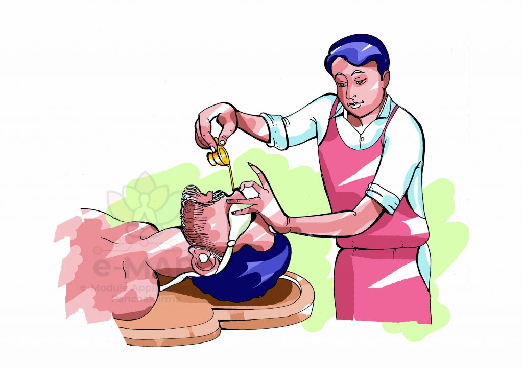
NASYA KARMA

EXPLANATORY NOTES
तद्ध्युत्तमाङ्गमनुप्रविश्य मुञ्जादीषिकामिवासक्तां केवलं विकारकरं दोषमपकर्षति|||२२||(Cha/Si/2/22)
According to Caraka, the NasyaDravya instilling into nostril will reach Uttamāṅga and expel the Doṣās there just like how Iṣikā grass has been removed from its stalk.
नासा हि शिरसो द्वारम्|
तत्रावसेचितमोउषधं स्रोतः-शृङ्गाटकं प्राप्य व्याप्य च मूर्धानं नेत्रश्रोत्रकण्ठादि-
सिरामुख़ानि च मुञ्जादिषीकामविसक्तामूर्ध्वजत्रुगतां
वैकारिकीमशेषामाशु दोषसंहतिमुत्तमाङ्गादपकर्षति| (As San/Su/29/3)
AshtaṅgaSangraha gave a detailed explanation that nose is the entry door to śiras. Hecne the medicine given through that route will fill all related Srotases and reach the ŚṛṅgāṭakaMarma and spread the whole of head and the channels and veins in the eyes, ears and throatby its Vīrya and expels out completely the accumulated vitiated Doṣās which are stationed in the parts above the clavicle just like an Iṣikā is removed from the Munja grass.
शृङ्गाटकमभिप्लाव्य निरेति वदनाद्यथा |(Su/Chi/40/29)
In nasyavyāpatSuśruta has told excessive VairecanikaNasya will result in MastulungaSrāva which shows the direct connection of Nāsā to Śiras.
According to the commentator Indu, the exact Sthāna of the ŚṛṅgāṭakaMarma is
“शिरसोअन्तर्मध्यम्मूर्धानां” which can be considered for the middle cranial fossa. On basis of above facts, the verse i.e. “नासाहिशिरसोद्वारं” can be justified which reflexes the action of Nasya in head and systemic disorders.
NASAL ANATOMY AND PHYSIOLOGY RELATED TO NASYA[i]
Nasal route runs from nasal vestibule to nasopharynx which has a depth of approximately 12 -15 cm. Total surface area of nasal cavity is about 180 sq.cmand it has total volume about 16-19 ml. Mucous lines this nasal route which protects the mucosa from the inspired air.
Nasal cavity is divided into three functional zones
- Vestibular region
- Respiratory region
- Olfactory region
Vestibular region: This region filters the air coming into the nasal cavity.
Respiratory region: This region carries out the function of respiration with maximum of its vascularity.
Olfactory region: Area of olfaction as well as act as connecting area between brain and vestibule.
The two nasal cavities are the uppermost part of the respiratory tract and contain the olfactory receptors. The smaller anterior region of the cavities is enclosed by the external nose whereas the large posterior region are more central within the skull. The openings of the para-nasal sinuses are the extension of the nasal cavity into the cranial cavity. Four para nasal sinuses are the ethamoidal, sphenoidal, maxillary and frontal sinuses. They develop as outgrowth from the nasal cavities and are lined by the mucous membrane. The nasal cavity is related with the anterior and middle cranial fossae, orbit and paranasal sinuses. The superior aspect of sphenoidal sinus is related with the hypophysis, the optic nerves, optic chiasma and literally to the cavernous sinus and internal carotid artery. The cribriform plate is a part of the ethmoid bone, forms the portion of the roof of the nasal cavity it contains very small perforations, allowing fibers of the olfactory nerve to enter and exit. Sphenopalatine foramen located at the level of superior meatus allows communication between the nasal cavity and the pterygo-palatine fossa.
The pharmacodynamics of nasyakarma can be explained in light of modern
anatomical and physiological studies as follows[i]:
- Vascular pathway: The nasal tissue is highly vascularized making it an
attractive site for rapid and efficient systemic absorption. Vascular path transportation is possible through the pooling of nasal venous blood into the facial vein which occursnaturally. It communicates freely with the intracranial circulation. Itcommunicates through pterygoid plexus with the cavernous venous sinus.
- Neurological pathway: Olfactory nerve is chemoreceptor in nature. It is known
that through olfactory pathway this nerve is connectedwith limbic system and hypothalamus which are having control over endocrine secretions.Moreover, hypothalamus is considered to be responsible forintegrating the functions of the endocrine system and the nervous system. So the drugs administratedhere stimulate the higher centers ofbrain which shows action on regulationof endocrine and nervous system functions.
- Diffusion through mucosa: In the absorption of drug from the nasal cavity
first step is passage through the mucus. Mechanisms for absorption of drug through the nasal mucosa include
- Paracellular route is the first mechanism which isan aqueous route of transport. This is slow and passive route.
- Transcellular process is the second mechanism of transport through a lipoidal route and is responsible for the transport of lipophilic drugs like snehanasyadravya that show a rate dependency on their lipophilicity. Drugs also cross cell membranes by an active transport route via carrier mediated means or transport through the opening of tight junctions.
These are the possible mode of action of how a nasyadravya works
[i]Sobiesk JL, Munakomi S. Anatomy, Head and Neck, Nasal Cavity. [Updated 2021 Jul 26]. In: StatPearls [Internet]. Treasure Island (FL): StatPearls Publishing; 2021 Jan-. Available from: https://www.ncbi.nlm.nih.gov/books/NBK544232/
[i]Kumar, V. (2017). A CONCEPTUAL STUDY ON MODE OF ACTION OF NASYA. International Journal of Ayurveda and Pharma Research, 5(7).
Blood Supply: Blood supply the nose has very rich vascular supply. It receives blood from both the internal and external carotid artery. Anterior and posterior ethmoidal artery are the branches of the internal carotid artery, descending to the nasal cavity through the cribriform plate. These arteries form anastomoses with each other in the anterior part of nose. The vein of the nose follow the arteries and they drain into the pterygoid plexus, facial vein or cavernous sinus. A few nasal veins join with the sagittal sinus that is a Dural venous sinus. This is a potential pathway through which infection may spread from nose to cranial cavity. The lymph vessel drains into deep cervical node. Communication probably occur between the nasal lymphatics and subarachnoid space, probably through the sheath of the olfactory nerve.
- Nerve Supply: In brief olfaction is provided by the olfactory nerve (CN I). General sensation is carried by the trigeminal nerve (CN V). Motor innervation to the nasal muscles of facial expression is via the facial nerve (CN VII) and serous glands in the nasal mucosa which produce fluid that constantly lubricates the nose walls are innervated by the parasympathetic fibers of the facial nerve (CN VII). Sympathetic innervation comes from T1 level of spinal cord and is intended for regulation of blood flow through mucosa. The lateral aspects of the nose are supplied by the infrorbital nerve, a branch of the maxillary nerve (CNV2). Olfactory nerve which provides a special sensory innervation through the cribriform plate. Olfactory bulb a part of the brain lies on the superior surface of the cribriform plate, above the nasal cavity.
The entire nasal cavity is covered by a special lining called mucosa. This mucosal surface has microscope hair like structure called cilia and many mucuous producing glands. The physiological functions of the nose include: respiration, conditioning the respired air, vocal resonance, olfaction and nasal resistance, protection of the lower airways, ventilation and drainage of the sinuses.
गुदापस्तम्भविधुरशृङ्गाटानि नवादिशेत्|
मर्माणि धमनीस्थानि—-(As Hr/Sa/4/40)
ŚṛṅgāṭakaMarma is a DhamaniMarma and SadyoPrāṇaharaMarma.
जिह्वाक्षिनासिकाश्रोत्रखचतुष्टयसङ्गमे|
तालुन्यास्यानि चत्वारि स्रोतसां, तेषु मर्मसु||३४||
विद्धः शॄङ्गाटकाख्येषु सद्यस्त्यजति जीवितम्| (As Hr/Sa/4/35)
Its the meeting point of tongue, eyes, nose and ears.
Suśrutahas described that the ŚṛṅgāṭakaMarmais a SirāMarma, situated at the site of union of siras, supplying to the nose, ears, eyes, tongue.
These are four in number, incorporated under SadyoPrāṇaharacategory, injury to these causes quick death.Measures vicinity of four Aṅgula(8cm) circumferences. The corpus of this marma is made up of Sirāor Dhamani (vascular entity). On injury of these Marma, death will occur immediately or within seven days . The Nasya medicaments are going to reach in the Śṛṅgāṭaka area and the Doṣaresiding in the region of oral cavity (Mukha), nose (nāsā), eye (Akṣi) & tongue (jihvā) are expelled out.
OLFACTORY NERVE AND CENTRES[i]
The olfactory nerve is the first of the 12 cranial nerves and one of the few cranial nerves that carries special sensory information only. In this case, the olfactory nerve is responsible for our sense of smell.
In olfaction ,olfactory nerve is just a component. Pathway can be summarised as follows:
- olfactory nerves
- olfactory bulb
- olfactory tract
- olfactory striae
- olfactory cortex
- output targets of the olfactory cortex
Olfactory Receptor Cells: These cells are located in the olfactory epithelium, a mucosal membrane that lines the roof and sides of the nasal cavity. the olfactory epithelium is small; approximately 5 cm² in area. There are three cell types contained within the epithelium: the olfactory receptor cells, supporting cells, and basal (stem) cells.
The olfactory receptor cells are bipolar, meaning that they have two projections from their cell body. One projection, the dendrite, extends to the surface of the olfactory epithelium. This dendrite expands at the epithelial surface to become knob-like. Located on the dendrite’s surface are 10-20 non motile cilia that extend into the fluid layer covering the epithelium in the nose. The cilia contain receptors for odor molecules that pass into the nasal cavity and are captured in the fluid covering the olfactory epithelium. The other projection from the receptor cell body is an unmyelinated axon.
Other cell type over there the basal stem cells differentiate into, and replace, damaged receptor cells.
Olfactory Nerve: Each receptor cell has an axon extending from its basal surface. The basal surface of olfactory receptor cells is located directly inferior to the cribriform plate of the ethmoid bone which makes up the bony roof of the nasal cavity. As the axons project from the cell body, they combine with other receptor cell axons, making up bundles of nerve fibers/rootlets. All of these axonal bundles can collectively be thought of as the olfactory nerve (CNI). These bundles of nerve fibers, surrounded by dura and arachnoid mater, then move superiorly by passing through the foramina (holes) in the cribriform plate of the ethmoid bone.
Olfactory Bulb:
The axons projecting from the olfactory receptor cells via the olfactory nerve terminate within the olfactory bulb. The olfactory bulb is the main relay station within the olfactory pathway. Information from the receptor cells is passed to cells whose projections make up the subsequent olfactory tract.Each olfactory bulb (right and left) lies lateral to the crista galli and superior to the cribriform plate of the ethmoid bone, inside the cranial cavity. Therefore, it lies on the underside of medial aspect of the frontal lobe. Within the olfactory bulb are bundles of nerve fibers known as glomeruli; where incoming receptor cell axons make connections with the dendrites of mitral relay neurons.
Olfactory Tract: Each olfactory bulb (right and left) lies lateral to the crista galli and superior to the cribriform plate of the ethmoid bone, inside the cranial cavity. Therefore, it lies on the underside of medial aspect of the frontal lobe. Within the olfactory bulb are bundles of nerve fibers known as glomeruli; where incoming receptor cell axons make connections with the dendrites of mitral relay neurons.
Olfactory Striae: Posterior and anterior to the optic chiasm, the olfactory tract on both sides divides into medial and lateral olfactory striae. The medial stria projects to the anterior commissure, and subsequently, to contralateral olfactory structures. The lateral stria continues on to structures associated with the olfactory cortex.
Olfactory cortex: This cortex is not a single structure, rather, it is defined as the combined areas of the cerebral cortex (generally within the temporal lobe) that receive input directly from the olfactory bulb. These regions include the:
- Piriform cortex: which is located below the lateral olfactory stria.
- Amygdala: which is located anterior to the temporal/inferior horn of the lateral ventricle and is associated with the emotion of fear.
- Entorhinal cortex: which is the anterior part of the parahippocampalgyrus, and is involved in the formation of memory.
Olfactory Cortex Output Structures:
- From the olfactory cortex, information about smell is sent to the orbitofrontal cortex via the dorsal medial nucleus of the thalamus. The orbitofrontal cortex is a portion of the prefrontal cortex that is located on the underside of the frontal lobe and situated over the eye orbit. Lesions of this cortical region can result in an inability to distinguish different odors. Odor information is also sent to portionsof hypothalamus and brainstem that trigger autonomic responses involved in appetite, salivation, and gastric contraction.
[i]https://www.kenhub.com/en/library/anatomy/the-olfactory-pathway


